Electromyography, or EMG, is a diagnostic procedure used to assess muscle function and the nerves controlling them (motor neurons). The results of this examination can reveal nerve disorders, muscle disorders, or issues with nerve transmission to the muscles.
Contents
When is Electromyography Recommended?
EMG is typically recommended by doctors when patients experience symptoms such as recurring tingling, numbness, muscle weakness, cramps, muscle pain, or discomfort in the legs or hands.
Some conditions that can be diagnosed using this examination include:
- Peripheral nerve disorders such as peripheral neuropathy and carpal tunnel syndrome
- Disorders affecting nerve endings in the spine, such as pinched nerves and sciatica
- Muscle disorders such as muscular dystrophy, polymyositis, and dermatomyositis
- Motor neuron disorders in the brain or spine, such as amyotrophic lateral sclerosis (ALS)
- Disorders affecting the connection between nerves and muscles, such as myasthenia gravis.
Electromyography Procedure
The EMG procedure is relatively straightforward. Here’s what to expect during the examination:
Before EMG
Preparation before an EMG is not complicated. You simply need to shower and wear comfortable clothing, avoiding the use of creams or lotions on your skin to ensure the accuracy of the examination.
If you regularly take blood-thinning medications or anticoagulants such as warfarin, inform your doctor. Your doctor will instruct you to stop taking them for several days to reduce the risk of bleeding during the procedure.
Also, inform your doctor if you use electrical medical devices such as pacemakers.
During EMG
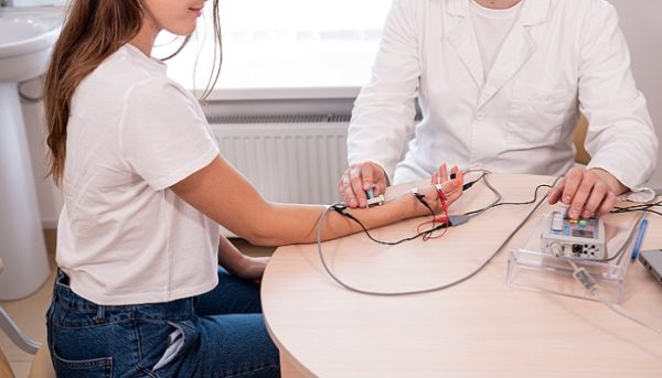
EMG begins after a procedure called nerve conduction study is conducted.
During the nerve conduction study, the doctor will place electrode stickers on the skin surface. These electrodes will emit mild electrical impulses that stimulate the nerves to function. The nerve responses will then be recorded by the device.
After the nerve conduction study, the EMG procedure will commence. Here’s what happens:
- You’ll be instructed to lie down on an examination bed.
- The doctor will identify the muscle location to be examined and insert a needle into that area.
- You’ll be asked to relax and move the muscle in specific ways to assess its function.
- These movements will be recorded by the device and displayed as graphs on the screen to show the electrical activity in the muscle.
- Once the necessary data is collected, the needle will be removed. This process may be repeated several times in different locations.
The procedure typically takes 60-90 minutes, depending on the number of areas being examined. Some patients may experience discomfort during the needle examination, but most can tolerate it without significant pain.
After EMG
Electromyography results are usually available within one or two days after the examination.
After the EMG procedure, you may experience soreness at the needle insertion site, which will subside within a few days. Bruising may also occur in the same area.
Although rare, this procedure carries risks of bleeding, severe pain, or infection, characterized by fever and swelling at the examination site.
If you’re experiencing symptoms of nerve disorders that are affecting you, promptly consult a neurology specialist. Mandaya Royal Hospital Puri houses a comprehensive nerve examination center with state-of-the-art equipment and experienced specialist doctors. Don’t hesitate to schedule your appointment with a doctor now through WhatsApp Chat, the Book Appointment page, or the Care Doctor app, available for download on Google Play and the App Store. In addition to appointments, you can monitor your queue number and access other relevant information there.