For individuals experiencing brain disorders, several diagnostic tests can be conducted, one of which is electroencephalography or EEG. This procedure aids in diagnosing conditions such as epilepsy, brain tumors, and encephalitis. EEG is performed by neurology specialists trained and skilled in operating the equipment.
Contents
What is EEG?
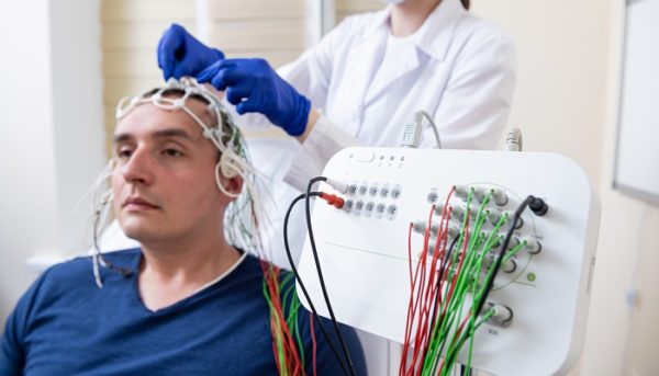
Electroencephalography (EEG) is a test used to measure electrical activity in the brain using electrode discs resembling coins attached to the scalp. These electrodes capture the electrical impulses emitted by brain cells, which are then displayed graphically on a screen.
EEG is most commonly used to diagnose epilepsy. However, this procedure can also be utilized to diagnose other brain disorders.
Why should be done?
EEG procedures are beneficial in detecting changes in brain activity. If any abnormal changes occur, doctors can diagnose the underlying cause of the disorder. Some brain diseases that can be detected using this examination include:
- Epilepsy
- Brain tumors
- Brain damage due to injury or accidents
- Brain function disorders or encephalopathy
- Sleep disorders
- Encephalitis
- Stroke
This examination can also be used to confirm brain death in coma patients. Sometimes, EEG is performed continuously, especially in medical coma patients, to determine the appropriate anesthesia dosage.
Electroencephalogram Procedure
Below are the steps involved in performing an EEG procedure:
Before EEG
Before undergoing EEG, you will be instructed to shampoo your hair up to two times. This is necessary to ensure that the scalp area is completely clean, allowing the equipment to be properly attached without disrupting the examination results.
You are also not allowed to use hair products such as gel or wax.
Your doctor will also advise you to get sufficient sleep the night before the examination, so you can fall asleep during EEG.
During EEG
- The doctor will measure your head and mark the locations where the electrodes will be placed.
- The marked areas will be applied with a special cream to help the electrodes obtain clearer readings.
- The doctor or nurse will then attach adhesive to the electrodes, which will be placed on the scalp.
- Once the examination begins, the electrodes will transmit electrical impulse data from the brain to the device and display it graphically on the screen.
- During the examination, the doctor may direct you to perform various tasks, such as taking deep breaths, examining stimuli by directing light into your eyes, and so on. This is done to observe different responses.
- The examination usually lasts for 30-60 minutes.
After EEG
After the procedure is completed, all equipment attached to the scalp will be removed. You can resume your daily activities immediately, although there may still be adhesive residue on your scalp, so you are usually advised to shampoo afterwards.
For certain patients who are anesthetized during the procedure, it is necessary to wait for the effects of anesthesia to wear off before resuming activities.
Neurology specialists will interpret the EEG results recorded in graphical form. Graphs of normal brain conditions will exhibit different patterns from those with specific disorders.
If you experience disruptive neurological symptoms, consult a neurology specialist. Mandaya Royal Hospital Puri has a neurological examination center equipped with newest technology and and experienced specialists.
Feel free to schedule an appointment with a doctor via our WhatsApp Chat, the Book Appointment page, or the Care Doctor app, available for download on Google Play and the App Store. In addition to appointments, you can also monitor your queue number and access other comprehensive information there.