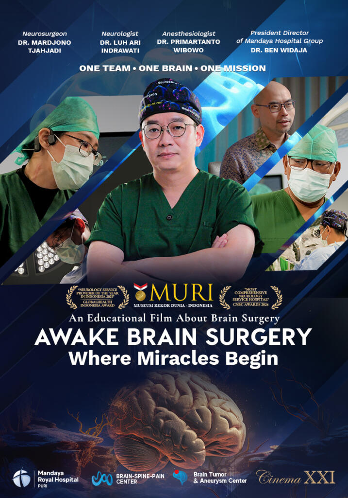
Brain tumors are serious conditions that require careful treatment, especially when the tumor is located near areas of the brain responsible for vital functions such as speech, movement, and vision. One surgical approach that is increasingly recognized for handling such cases is awake brain surgery, a brain operation in which the patient remains conscious during certain stages of the procedure. This technique allows the surgical team to monitor brain function directly throughout the operation, helping protect critical areas from damage.

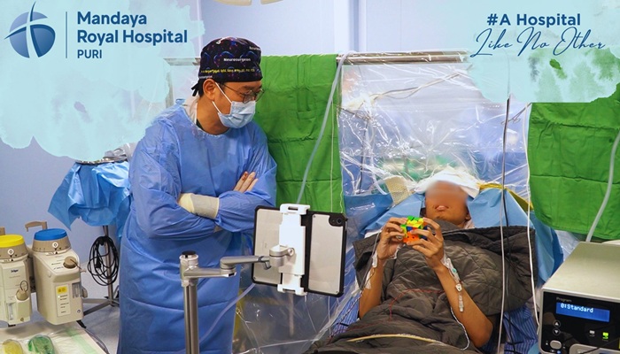
Recently, a brain tumor patient underwent awake brain surgery with Dr. dr. Mardjono Joy Tjahjadi, Sp.BS, Subsp. N-Vas, F. N-Onk, PhD, FICS, IFAANS, better known as Dr. Joy, at Mandaya Royal Puri Hospital. What made this case unique was that the patient played with a Rubik’s Cube during the surgery and was even able to solve it successfully.

Contents
Tumor Successfully Removed 100%, Patient Walked Immediately After Surgery
The awake brain surgery performed by Dr. Joy at Mandaya Royal Puri Hospital was completed safely and successfully. The patient’s brain tumor was entirely removed—100%—without significant complications. This success demonstrates that the awake brain surgery approach is not only effective but also capable of delivering optimal outcomes, even for tumors located in high-risk areas.
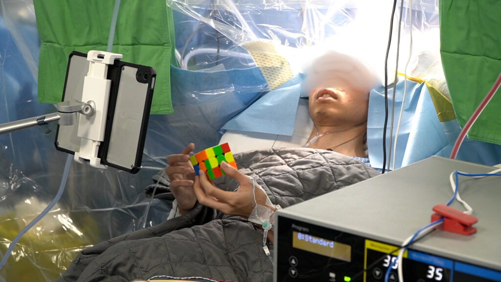
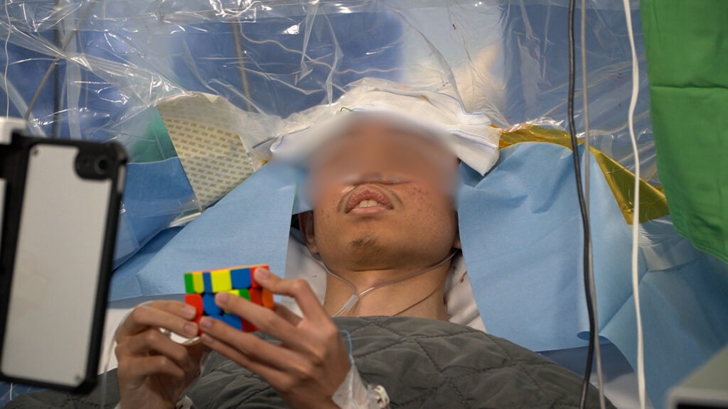
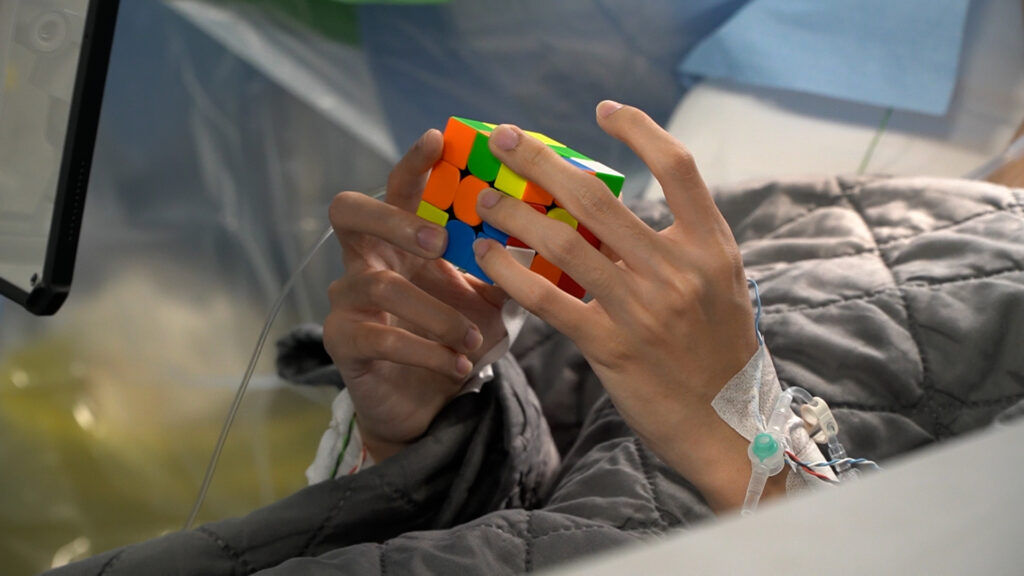
During the surgery, the patient was asked to play with a Rubik’s Cube as part of real-time brain function monitoring, ensuring that the areas controlling motor and cognitive functions remained unaffected while the tumor was being removed. What made the moment even more remarkable was that Dr. Joy and the patient held a small “competition”: who would finish first—the patient solving the Rubik’s Cube or Dr. Joy removing the tumor?
In the end, the patient solved the Rubik’s Cube first, and about a minute later, the tumor was completely removed.
Immediately after the tumor was extracted, Dr. Joy showed the tumor directly to the patient. This became a clear sign that the operation had been completed successfully and according to plan, while the patient—still fully conscious—was able to witness the moment firsthand.
Not only that, shortly after the procedure ended, the patient was able to walk immediately and was guided by Dr. Joy to step down from the operating table. This rapid recovery showed that the patient’s motor functions remained well-preserved throughout the surgery.
What Is Awake Brain Surgery and Why Is It Needed?
Awake brain surgery, also known as awake craniotomy, is a brain surgery procedure in which the patient remains conscious during the operation. While the patient is awake, the medical team can monitor brain function directly and in real time. This procedure is most commonly used when the treated area is close to parts of the brain that control important functions such as speech, movement, and vision.
So, why is this method necessary in certain cases? Here is the explanation:
-
High-Risk Tumor Location
When a tumor or seizure-causing area is located near parts of the brain responsible for critical functions such as speaking, moving, or comprehension, surgeons need to remove the tumor while preserving those vital abilities.
-
Imaging Alone Is Not Enough
Scans such as MRI can show the tumor’s location, but they cannot always precisely identify where important functions are located in every individual. These functional areas may vary from person to person, especially if the tumor or seizures have altered brain activity.
-
Real-Time Brain Function Monitoring
With the patient awake, the surgical team can assess brain function in real time by asking the patient to speak, move, or respond to stimuli, allowing important areas to be identified and protected during surgery.
-
Brain Mapping for Safer Navigation
Brain mapping techniques are used to identify areas of the brain responsible for language, movement, sensation, and vision. This method is extremely useful when operating near critical functional centers.
-
More Optimal Outcomes
This approach helps surgeons remove as much tumor tissue or seizure-causing tissue as possible while minimizing the risk of impairments to speech, movement, or other important functions after surgery.
Brain Tumor Surgery Becomes Safer with IONM Technology at Mandaya Puri Hospital
The success of this awake brain surgery procedure was also supported by the availability of Intraoperative Neuromonitoring (IONM) technology at Mandaya Royal Puri Hospital. IONM is a real-time nerve function monitoring system used during surgery, allowing the surgical team to detect early changes in brain, spinal cord, and peripheral nerve function to help prevent permanent damage.
With IONM, the surgical team receives an “early warning” if activity in certain nerve areas begins to show unwanted changes during the procedure. This allows Dr. Joy and the team to immediately adjust their surgical techniques in real time, significantly reducing the risk of impairments to speech, movement, and other neurological functions.
The presence of this technology positions Mandaya Royal Puri Hospital as one of the hospitals in Indonesia capable of providing neurosurgical safety standards comparable to international facilities, offering greater peace of mind for patients and families facing even the most complex brain surgeries.
Dr. Joy’s Profile and Practice Schedule at Mandaya Royal Puri Hospital

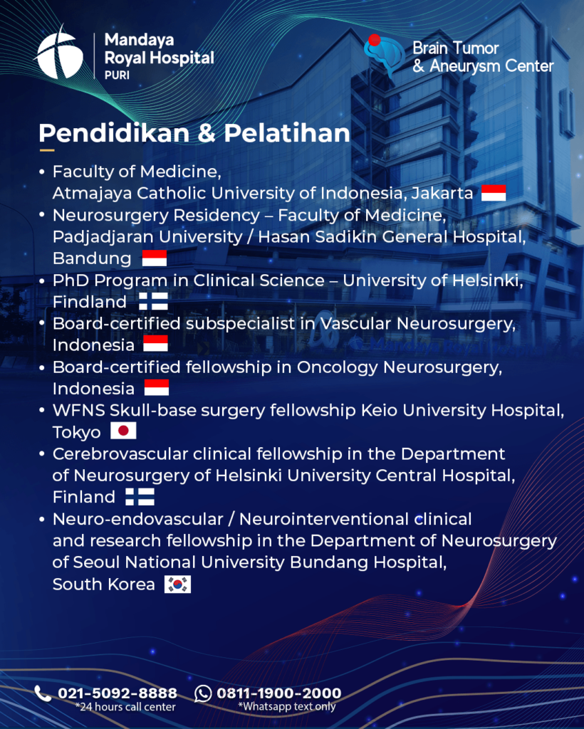
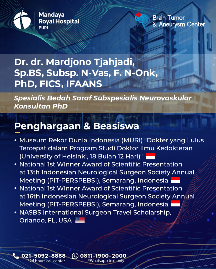
Dr. dr. Mardjono Joy Tjahjadi, Sp.BS, Subsp. N-Vas, F. N-Onk, PhD, FICS, IFAANS, commonly known as Dr. Joy, began his medical journey at one of Indonesia’s leading universities, where his passion for neurosurgery and neurovascular treatment first developed. He later continued his neurosurgery specialization training in Finland, where he learned directly from world-class neurosurgeons while actively participating in cutting-edge research on brain aneurysms and brain tumors.
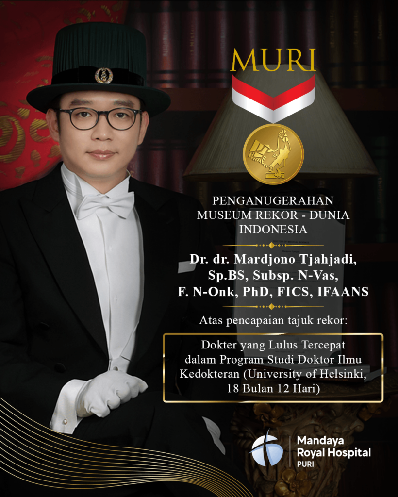
One of the most remarkable achievements in his academic career was completing his doctoral degree (PhD) in Finland in just 18 months and 12 days, earning him an Indonesian World Records Museum (MURI) award as the “Fastest Doctor to Complete a Medical PhD.”
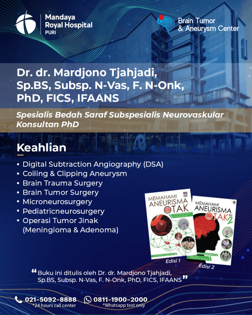
Today, Dr. Joy is recognized as one of Indonesia’s leading neurosurgeons, with special expertise in treating brain aneurysms, brain hemorrhages, brain tumors, and brain cancer. His strong educational background combined with extensive international experience has earned him numerous national and international awards.
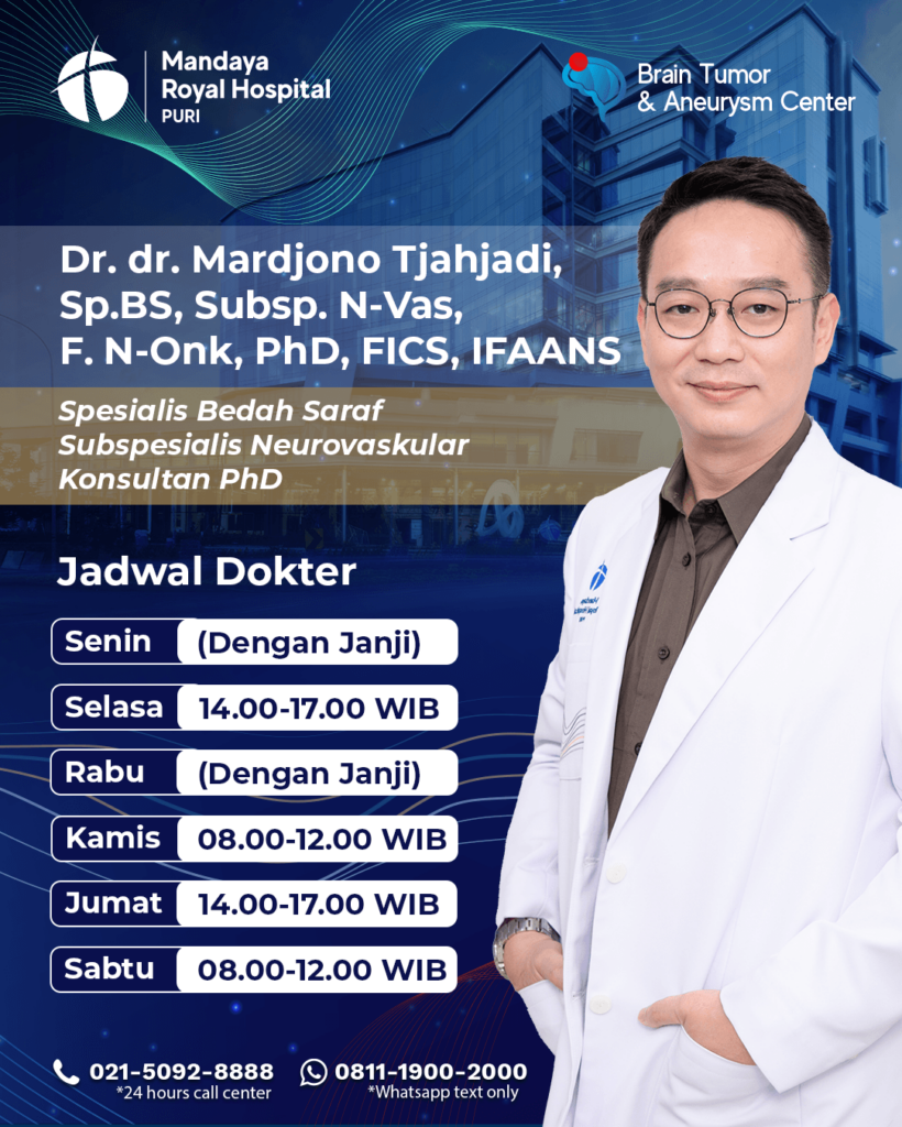
Dr. Joy’s Practice Schedule at Mandaya Royal Puri Hospital
- Monday: By appointment
- Tuesday: 2:00 PM – 5:00 PM WIB
- Wednesday: By appointment
- Thursday: 8:00 AM – 12:00 PM WIB
- Friday: 2:00 PM – 5:00 PM WIB
- Saturday: 8:00 AM – 12:00 PM WIB
To make your visit to Mandaya Royal Hospital Puri more convenient, you can use the WhatsApp Chat feature, Book Appointment service, or the Care Dokter application, available on Google Play and the App Store, to simplify appointments, check queue numbers, and access other complete information.
The information provided on this page is intended for educational and general informational purposes only and does not reflect the full range of medical services that may be performed by each doctor. To ensure treatment appropriate to your medical condition, direct consultation with the relevant doctor is recommended.
If you have any questions, suggestions, or need further information, please contact our call center at 0811-1900-2000.