Pancreatic cancer is one of the most challenging types of cancer to treat. In addition to its symptoms often going undetected in the early stages, this cancer is frequently diagnosed at an advanced stage, where the tumor has already involved surrounding blood vessels or organs, making surgical removal impossible.
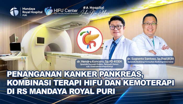
However, advances in medical technology are now opening new possibilities. At Mandaya Royal Puri Hospital, a multidisciplinary approach combining chemotherapy with HIFU (High Intensity Focused Ultrasound) technology offers a non-invasive solution for patients who are unable to undergo surgery.

Contents
Patient Condition and Treatment Challenges
The patient in this case was a 78-year-old elderly individual diagnosed with advanced-stage pancreatic cancer. A tumor measuring approximately 4 cm was found in the body (corpus) of the pancreas. After undergoing four cycles of chemotherapy, the tumor shrank to around 3 cm with a diameter height of 2.7 cm.
Nevertheless, the tumor’s condition still did not allow surgery to be performed. As explained by dr. Sugianto Santoso, Sp.Rad (K)RI, an interventional radiology specialist at Mandaya Royal Puri Hospital, the tumor had completely encased the common hepatic artery and splenic artery while also involving part of the duodenal wall. The extensive involvement of these major blood vessels became the main obstacle preventing surgical intervention.
Therefore, the medical team at Mandaya Royal Puri Hospital decided to combine chemotherapy with the HIFU procedure, a technology that can be used for patients who are not eligible for surgery.
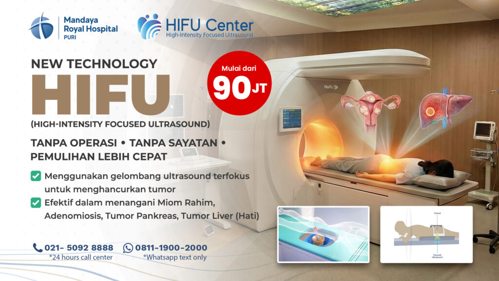
HIFU is a non-invasive therapy that uses high-intensity sound waves to generate extreme heat at a targeted focal point. In pancreatic cancer cases, this technology functions as a thermal ablation therapy, destroying tumor tissue without the need for surgical procedures or incisions.
Here are some of the advantages of HIFU for pancreatic cancer:
- No surgery or incisions: Minimizes the risks of bleeding, infection, and physical trauma.
- Pain relief: Proven effective in reducing pain intensity, especially in advanced-stage patients.
- Tumor shrinkage: Breaks down tumor masses without damaging surrounding healthy tissue.
- Lower risk: Better tolerated and suitable for patients who cannot undergo major surgery.
- No radiation: Uses sound waves, making it safe to repeat if necessary.
Diagnostic Process: Endoscopic Ultrasound Biopsy (EUS) as the Gold Standard
Before determining the treatment strategy, establishing an accurate diagnosis was a crucial first step.
dr. Hendra Koncoro, Sp.PD-KGEH, an internal medicine specialist and consultant in gastroenterology-hepatology at Mandaya Royal Puri Hospital, explained that the procedure used was EUS-FNA (Endoscopic Ultrasound Fine Needle Aspiration), an endoscopic ultrasound biopsy considered the gold standard for pancreatic cancer evaluation.
In this procedure, an endoscope equipped with ultrasound is inserted through the mouth until it reaches the duodenum. From there, doctors can directly access the pancreas, perform imaging, and collect tissue samples using a fine needle for laboratory examination.
The results of this examination help doctors determine the cancer stage as well as the type of cells present, providing essential information for selecting the most appropriate therapy.
According to dr. Hendra, in early-stage cases (stage 1) where the tumor is smaller than 2 cm and has not spread, surgery is still possible and may provide good remission outcomes. However, when the tumor has progressed to an advanced stage and involves blood vessels, treatment options shift toward chemotherapy, immunotherapy, or ablative procedures such as HIFU.
HIFU Technology as a New “Weapon” in Combination Therapy
After completing four cycles of chemotherapy, dr. Sugianto and dr. Hendra proceeded with the HIFU procedure. This technology works by focusing high-energy ultrasound waves precisely on the tumor site, allowing cancer tissue to be destroyed without incisions or surgery.
The success of HIFU in this patient was strongly supported by pre-procedure simulation results: the patient’s stomach did not obstruct the pancreatic or tumor area, allowing the ultrasound waves to reach the entire tumor mass without interference. This made the patient an ideal candidate for the HIFU procedure.
After the procedure, the patient will be evaluated within 1.5 to 2 months. If residual tumor tissue remains and surgery is still not possible, the medical team may continue chemotherapy or consider other ablative techniques such as cryoablation if the patient’s condition allows.
This multidisciplinary approach, supported by advanced technologies such as HIFU at Mandaya Royal Puri Hospital, offers new hope for patients with advanced-stage pancreatic cancer—not only to shrink tumors but also to improve the patient’s overall quality of life.
Consultation with dr. Sugianto and dr. Hendra Koncoro at Mandaya Royal Puri Hospital
The success of pancreatic cancer treatment at Mandaya Royal Puri Hospital is inseparable from the role of highly experienced specialists in their respective fields. Below are the profiles of the two doctors directly involved in this case.
1. dr. Hendra Koncoro, Sp.PD-KGEH

dr. Hendra Koncoro, Sp.PD-KGEH is an internal medicine specialist and gastroenterohepatology consultant at Mandaya Royal Puri Hospital. He is experienced in treating various digestive tract and liver conditions, including liver cancer, gallstones, fatty liver disease, liver nodules, liver cirrhosis, and bile duct cancer.
He completed his general medical education at Atma Jaya Catholic University, followed by a Master’s degree in Biomedical Sciences and Internal Medicine Specialist training at Udayana University, before completing his Gastroenterology & Hepatology Consultant training at the University of Indonesia.
dr. Hendra Koncoro, Sp.PD-KGEH is available at Mandaya Royal Puri Hospital on:
- Wednesday: 10:30 AM – 12:30 PM WIB
- Saturday: 10:30 AM – 12:30 PM WIB
2. dr. Sugianto Santoso, Sp.Rad (K)RI

dr. Sugianto Santoso, Sp.Rad (K)RI is a radiology specialist with a subspecialty in interventional radiology at Mandaya Royal Puri Hospital. He focuses on minimally invasive procedures for diagnosing and treating diseases in various organs using radiology imaging guidance. His expertise includes TACE, RF Ablation, and Microwave Ablation for treating liver nodules and liver cancer.
He completed his general medical education at Maranatha Christian University Bandung before continuing his Radiology Specialist training at the University of Indonesia.
dr. Sugianto Santoso, Sp.Rad, (K)RI is available at Mandaya Royal Hospital Puri on:
- Tuesday: 3:00 PM – 6:00 PM WIB
- Thursday: 12:00 PM – 4:00 PM WIB
- Saturday: 10:00 AM – 1:00 PM WIB.
To make your visit to Mandaya Royal Hospital Puri more convenient, you can use the WhatsApp Chat feature, Book Appointment service, or the Care Dokter application, available on Google Play and the App Store, to simplify appointments, check queue numbers, and access other complete information.
The information provided on this page is intended for educational and general informational purposes only and does not reflect the full range of medical services that may be performed by each doctor. To ensure treatment appropriate to your medical condition, direct consultation with the relevant doctor is recommended.
If you have any questions, suggestions, or need further information, please contact our call center at 0811-1900-2000.