Gastroschisis is a congenital birth defect in which a baby’s intestines (stomach, large intestine, or small intestine) protrude outside the body through an opening about 2–5 centimeters wide, usually located beside the belly button, during fetal development.
This condition occurs early in pregnancy when the abdominal wall does not form properly, leaving a gap that allows the organs to slip outside the body. These exposed organs are then bathed directly in amniotic fluid, which may cause irritation and swelling. To treat the condition, the baby requires immediate surgery after birth to return the organs into the abdominal cavity.
Contents
Gastroschisis vs. Omphalocele
Both gastroschisis and omphalocele are congenital conditions where abdominal organs are located outside the body at birth. The main difference lies in the protective membrane:
- In omphalocele, the protruding organs are covered by a membrane.
- In gastroschisis, the organs are completely exposed without any protective covering.
This difference can usually be detected through prenatal ultrasound, allowing doctors to make an early diagnosis and prepare the right treatment plan.
What Causes Gastroschisis?
Gastroschisis occurs because the baby’s abdominal wall fails to form properly during pregnancy. The exact cause remains unknown. If you are planning a pregnancy, it is recommended to consult a healthcare provider to discuss ways to minimize the risk of congenital conditions in your baby.
Symptoms of Gastroschisis
During pregnancy, mothers typically do not experience direct symptoms of gastroschisis. However, prenatal ultrasound may reveal signs such as:
- Stomach, small intestine, or large intestine outside the baby’s body.
- Swollen intestines.
- Twisting or malrotation of the intestines.
- Low body temperature (hypothermia) in the baby.
Complications of Gastroschisis
Babies born with gastroschisis may face several health challenges, including:
- Premature birth: Babies with gastroschisis are at higher risk of being born early.
- Intestinal blockage: After surgery, the intestines may narrow, preventing food and waste from passing through smoothly.
- Short bowel syndrome: In rare cases, parts of the intestines may be underdeveloped or missing, leading to difficulty absorbing nutrients.
Diagnosis of Gastroschisis
Gastroschisis can be diagnosed both during pregnancy and after birth. Most cases are identified at 18–20 weeks of pregnancy during routine prenatal screenings for birth defects. Diagnostic methods may include:
- Ultrasound (USG): Imaging that uses sound waves to view soft tissues inside the body.
- Blood tests: Specifically measuring levels of alpha-fetoprotein (AFP). Elevated AFP between weeks 18–22 of pregnancy may indicate gastroschisis.
- MRI (Magnetic Resonance Imaging): Provides detailed images of the mother’s and baby’s internal structures.
Treatment for Gastroschisis
Although gastroschisis can be detected during pregnancy, treatment is only possible after the baby is born.
The main treatment is surgery to return the organs into the abdominal cavity and close the abdominal opening near the belly button. The type of surgery depends on the severity and the number of organs outside the body:
- Primary repair: If possible, the baby undergoes surgery immediately after birth to return the organs inside and repair the abdominal wall.
- Staged repair: For more complex cases, surgery is performed in stages. This is often chosen when the baby is too weak for a major operation or when the abdominal cavity cannot accommodate all organs at once.
Before surgery, the exposed organs are placed in a sterile plastic pouch called a silo to protect them from infection, dehydration, and damage. After the initial repair, additional surgeries may be needed to strengthen the abdominal muscles or intestines.
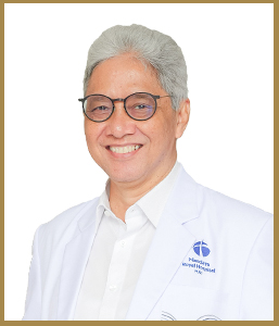
Pediatric Surgery Expert at Mandaya Royal Hospital Puri

Mandaya Royal Hospital Puri is home to one of Indonesia’s leading pediatric surgeons, dr. Sastiono, Sp.B Subsp.Ped(K). He is widely recognized for his expertise in managing rare and complex pediatric surgical conditions, including gastroschisis.
dr. Sastiono completed his medical training at the Faculty of Medicine, University of Indonesia (FK UI), progressing from general surgical training to pediatric surgery specialization, and later becoming a Consultant in Pediatric Surgery. He is highly skilled in pediatric hepatobiliary surgery and liver transplantation, covering surgical procedures on the liver, gallbladder, bile ducts, and pancreas.
In addition to gastroschisis and omphalocele, dr. Sastiono is experienced in treating various pediatric surgical conditions, such as:
- Biliary atresia (blockage of bile ducts in infants).
- Anal atresia (congenital malformation of the anus).
- Pediatric hernia.
- Appendicitis.
- Intestinal obstruction.
- Pediatric liver disorders.
- Hirschsprung disease.
With his comprehensive expertise, dr. Sastiono has become a trusted referral for parents seeking safe, professional, and reliable pediatric surgical care.
dr. Sastiono’s Practice Schedule at Mandaya Royal Hospital Puri
- Tuesday: 10:00 – 13:00 WIB
- Thursday: 10:00 – 13:00 WIB
If you wish to consult with dr. Sastiono regarding gastroschisis or other pediatric surgical conditions, you can visit Mandaya Royal Hospital Puri.
For easier access, use the WhatsApp Chat feature, Book Appointment, or the Care Dokter app (available on Google Play and App Store) to schedule visits, check queue numbers, and obtain complete hospital information.


