After completing cancer treatment, patients still need follow-up tests to check whether there are any remaining cancer cells in the body. While cancer treatment is designed to eliminate cancer cells, some may persist, requiring further treatment to remove them completely.
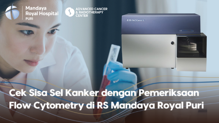
To detect residual cancer cells, Mandaya Royal Puri Hospital provides flow cytometry testing.

Contents
What is Flow Cytometry?
Flow cytometry is a laboratory test used to analyze the characteristics of cells or particles.
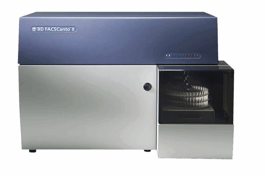
During the procedure, cells are stained with fluorescent antibody dyes to mark specific proteins on the surface or inside the cells. These cells are then passed through a flow cytometer, which can process and analyze around 10,000 cells in less than one minute.
Flow cytometry can be used for:
- Counting cells
- Sorting cells
- Determining cell characteristics and functions
- Diagnosing and treating blood cancers or bone marrow cancers
- Identifying biomarkers (measurable substances that predict cancer behavior or treatment response)
- Immunophenotyping (classifying types of cells)
When is Flow Cytometry Needed?
Doctors may recommend flow cytometry after a cancer diagnosis. This test helps determine the right time to start treatment and whether the treatment is effective.
Once cancer treatment is completed, flow cytometry can be part of a patient’s routine follow-up testing to detect if the cancer has returned.
This test is especially valuable for identifying minimal residual disease (MRD)—small numbers of cancer cells that remain after treatment. Flow cytometry can detect as few as one cancer cell among 1,000 to 10,000 cells. Detecting MRD is crucial for deciding whether patients need further treatment or should try a different type of cancer therapy.
How Flow Cytometry Works
Flow cytometry quickly analyzes the physical and chemical properties of cells and particles. The process involves:
- Laboratory staff place a patient’s blood, tissue, or bone marrow sample in liquid, stain it with fluorescent antibody dyes, and inject it into the flow cytometer.
- The flow cytometer arranges cells in a single stream and passes them in front of a laser beam.
- The machine counts and categorizes cells based on how the laser light scatters off them.
- Once data is collected, the flow cytometer sends the results to a computer.
- The computer generates a report, usually in the form of dot plots or bar charts.
- A pathologist reviews the report and provides detailed findings in the test results.
Afterward, the doctor reviews the flow cytometry results along with the patient’s medical history, physical exam, and symptoms. They then explain whether the patient may have a disease such as cancer, discuss the meaning of the test results, and guide the patient on the next steps. Treatment options and personalized recommendations will also be provided.
Types of Cancer Detectable by Flow Cytometry
Flow cytometry can help detect several types of cancers, including:
- Acute lymphoblastic leukemia (ALL)
- Chronic lymphocytic leukemia (CLL)
- Non-Hodgkin lymphoma
- Acute myeloid leukemia (AML)
- Multiple myeloma
If you would like to consult about flow cytometry or get a referral for testing, visit Mandaya Royal Puri Hospital.
For your convenience, you can use the WhatsApp Chat, Book Appointment, or the Care Dokter app (available on Google Play and App Store) to manage your visit, check queue numbers, and access complete information.