fbpx

Coronary Heart Disease

What is Coronary Heart Disease?

Coronary heart disease is a condition that occurs when the heart’s arteries cannot deliver enough oxygen-rich blood to the heart muscle due to narrowing caused by fat deposits (plaque). This condition is also known as coronary artery disease or ischemic heart disease.

The coronary arteries are responsible for delivering blood to the heart muscle. If these arteries are damaged or diseased and unable to deliver oxygen and nutrients to the heart, it can lead to heart attacks and even death.

Symptoms of Coronary Heart Disease

The main symptoms of coronary heart disease include:

  • Chest pain (angina)
  • Shortness of breath
  • Pain in the neck, shoulders, jaw, or arms
  • Feeling faint
  • Nausea

Not everyone with coronary heart disease experiences the same symptoms. In fact, some people may not experience any symptoms at all before being diagnosed by a doctor.

Causes of Coronary Heart Disease

Coronary heart disease is caused by the buildup of fat, cholesterol, and other substances in and on the walls of the heart’s arteries. This condition is called atherosclerosis, and the deposits are known as plaque.

Plaque can cause arteries to narrow, blocking blood flow. Plaque can also rupture, leading to blood clots.

Some common causes of atherosclerosis and coronary heart disease include:

  • Diabetes or insulin resistance
  • High blood pressure (hypertension)
  • Lack of physical activity
  • Smoking or tobacco use

Diagnosis of Coronary Heart Disease

If a doctor suspects you are at risk for coronary heart disease, they may perform a risk assessment. The doctor will also ask about your personal and family medical history and lifestyle, and may order blood tests.

Further tests may be required to confirm a diagnosis of coronary heart disease, such as:

  • Electrocardiogram (ECG)
  • Echocardiogram
  • Chest X-ray
  • Coronary angiogram

Treatment for Coronary Heart Disease

1. Managing Risk Factors

There are several risk factors for coronary heart disease that you can control to prevent its onset, such as quitting smoking, managing cholesterol and blood sugar levels, improving your diet, maintaining an ideal weight, and managing blood pressure.

2. Medications

Doctors may prescribe medications for coronary heart disease, including:

  • Antiplatelets: Medications that reduce blood clotting, such as aspirin, clopidogrel, ticlopidine, and prasugrel.
  • Antihyperlipidemics: Medications to reduce blood lipids (fats), especially low-density lipoprotein (LDL) cholesterol.
  • Antihypertensives: Medications to lower blood pressure.

3. Coronary Angioplasty

Coronary angioplasty uses a balloon to widen the blood vessels, improving blood flow. Though angioplasty may be done on blood vessels in other parts of the body, it can help increase blood flow to the heart.

4. Heart Bypass Surgery

Heart bypass surgery is often performed on people who experience chest pain and have coronary heart disease. During the surgery, a bypass is created by grafting a vein above and below the blocked coronary artery area, allowing blood to flow around the blockage.

The vein is usually taken from the leg, but arteries from the chest and arms can also be used for the bypass graft. Sometimes, multiple bypasses are needed to restore full blood flow to all parts of the heart.

5. Rotablator

A rotablator is a device used to shave off hardened plaque in the coronary blood vessels, especially in cases where plaque is difficult to treat with balloons or stents.

Using a rotablator, doctors can remove blockages and improve or restore blood flow to the heart.

Want to consult further about coronary heart disease or overall heart health? Visit Mandaya Royal Hospital Puri. Our hospital is equipped with modern medical technology to handle various heart conditions. You can also consult with experienced heart specialists.

Schedule your appointment through our WhatsApp Chat feature, Book Appointment, or the Care Dokter app—available on Google Play and the App Store—to ease your visit, check your queue number, and access complete information.
Cholecystitis

What is Cholecystitis?

Cholecystitis is an inflammation of the gallbladder, a small, pear-shaped organ located beneath the liver that stores bile produced by the liver. Infection or blockage in the gallbladder or bile ducts can cause inflammation, pain, and swelling.

If left untreated, cholecystitis can lead to serious complications, such as gallbladder rupture, which can be life-threatening. Treatment often involves surgery to remove the gallbladder.

Symptoms of Cholecystitis

Common symptoms of cholecystitis include:

  • Severe pain in the upper right or middle abdomen
  • Pain radiating to the right shoulder or back
  • Tenderness in the abdomen when touched
  • Nausea
  • Vomiting
  • Fever

These symptoms often occur after eating, particularly fatty or large meals.

Causes of Cholecystitis

Several factors can cause gallbladder inflammation, including:

  • Gallstones Gallstones are the most common cause of cholecystitis. These hard particles form in the gallbladder and can block the duct that carries bile out of the gallbladder, leading to bile accumulation, swelling, and irritation.
  • Tumors A tumor can block the normal flow of bile from the gallbladder, leading to fluid buildup and inflammation.
  • Bile Duct Blockage A blocked bile duct can cause gallbladder inflammation. Scarring or bending of the bile ducts can also contribute to obstruction.
  • Infections Conditions like AIDS or other viral infections can cause gallbladder swelling and irritation.
  • Severe Illness Serious illnesses can damage blood vessels and reduce blood flow to the gallbladder, leading to inflammation.

Risk Factors for Cholecystitis

Certain individuals are at a higher risk of developing cholecystitis, including those who:

  • Are female
  • Are pregnant
  • Are obese
  • Have diabetes
  • Have high cholesterol levels
  • Experience rapid weight loss
  • Are over the age of 40

Diagnosis of Cholecystitis

To diagnose cholecystitis, doctors perform a physical examination and assess the patient’s symptoms and medical history. Diagnostic tests and procedures may include:

  • Blood Tests These tests check for signs of infection or other gallbladder problems.
  • Imaging Tests Imaging tests such as endoscopic ultrasound, abdominal ultrasound, CT scan, and magnetic resonance cholangiopancreatography (MRCP) provide images of the gallbladder and bile ducts, helping doctors identify signs of cholecystitis or gallstones.
  • Hepatobiliary Iminodiacetic Acid (HIDA) Scan This scan tracks the production and flow of bile from the liver to the small intestine. A radioactive dye is injected, which attaches to bile-producing cells, allowing doctors to detect any blockages.

Treatment for Cholecystitis 

Treatment options for cholecystitis include:

  • Fasting Patients may need to refrain from eating or drinking initially to relieve stress on the inflamed gallbladder.
  • Intravenous Fluids IV fluids help prevent dehydration.
  • Antibiotics Antibiotic medications may be necessary if the gallbladder is infected.
  • Pain Relievers Pain management medications help reduce discomfort and inflammation.
  • Gallbladder Drainage In some cases, a gallbladder drainage procedure (cholecystostomy) may be required to remove infection, especially for patients who cannot undergo gallbladder removal surgery.

Cervical Cancer

Cervical cancer is the third most common type of cancer among Indonesians. However, this cancer can be prevented with the HPV vaccine starting at the age of 12. Also known as cervical cancer, it can be cured or go into remission if treatment begins at an early stage.

What is cervical cancer?

Cervical cancer is the growth of uncontrollable abnormal cells that form a tumor on the cervix or neck of the uterus. The cervix is the area that connects the vagina to the uterus.

Causes

Almost 99% of cervical cancer cases are caused by HPV (Human Papillomavirus) infection. This is a very common type of virus that is often transmitted through sexual intercourse.

It is estimated that 8 out of 10 women may be infected with HPV in their lifetime, but most do not develop cervical cancer.

Most HPV infections do not cause symptoms and clear up on their own. However, in a small percentage of people, certain types of HPV can persist for years. It is this long-term HPV infection that can trigger cervical cancer in women.

In addition to HPV infection, your risk of this disease may also increase if you:

  • Are infected with HPV and use birth control pills for five years or more.
  • Are an active or passive smoker.
  • Have a weakened immune system.

Symptoms 

The most common symptoms of cervical cancer include:

  • Vaginal bleeding outside of menstruation
  • Unusual discharge, such as increased volume, a foul odor, or a different color than usual.
  • Pain or discomfort during sexual intercourse
  • Pelvic pain
  • Longer or heavier menstruation than usual
  • Bleeding after menopause

In the early stages of development, cervical cancer may not cause any specific signs and symptoms. In fact, not everyone diagnosed will experience symptoms until the cancer reaches advanced stages.

Diagnosis 

To detect the presence of cervical cancer, doctors may perform several tests, such as:

1. Pap Smear

A Pap smear is performed by taking a sample of cervical tissue to be examined in a laboratory for the presence of abnormal cells.

In addition to detecting cancer cells, this test can also detect cells that have the potential to become cancerous.

2. Biopsy

A biopsy is a more in-depth examination of cervical tissue to confirm the presence or absence of cancer cells in the cervix.

A biopsy can be performed during a colposcopy examination. Colposcopy is a procedure performed by a doctor using a magnifying instrument to examine the symptoms and signs of cancer in the cervix.

3. Imaging Tests

Imaging tests such as MRI and CT scans may also be performed to determine the presence of malignant tumors in the cervix. These tests are usually performed to determine the severity of cancer or to determine the stage of cancer.

Treatment 

Here are some types of treatment that can be done to treat cervical cancer:

1. Surgery

For early-stage cervical cancer that has not spread, treatment with surgery may be performed. Types of surgery include:

  • Cone biopsy or conization
    A procedure usually chosen if the tumor is still very small. It involves removing all cancerous tissue in the form of a triangle or cone and leaving other healthy cervical tissue intact. It is usually also chosen for individuals who plan to become pregnant in the future.
  • Trachelectomy
    In this procedure, the entire cervix and surrounding tissue are removed. The uterus is not removed in this method, so it can still be performed on individuals planning to become pregnant.
  • Hysterectomy
    Surgery to remove the cervix, uterus, part of the vagina, and tissue and lymph nodes around the cervix. This method is chosen if there are no plans for future pregnancy and is an effective step in preventing the recurrence of cervical cancer.

2. Radiation Therapy

Radiation therapy is a treatment that uses high-energy rays to kill cancer cells. Radiation can be performed externally, internally, or a combination of both.

  • External radiation: High-energy radiation beams are directed directly from outside to the affected area of cancer.
  • Internal radiation: Also called brachytherapy. The doctor will place a radiation source inside the cervix.

3. Chemotherapy

Chemotherapy involves the use of powerful drugs to kill cancer cells. Low-dose chemotherapy is often combined with radiation therapy to treat cancer that has not spread.

If you need screening or treatment for cervical cancer, you can visit the Cancer Center at Royal Mandaya Hospital, which is equipped with world-class cancer treatment technology.

The oncology genomic laboratory at Mandaya Royal Hospital Puri can accurately determine your cancer type and subtype so that you can receive the appropriate treatment.

Consult with a specialist now and get the best treatment for your cancer. Use the Chat feature via Whatsapp, Book Appointment, or the Care Doctor app which can be downloaded from Google Play and the App Store to facilitate visits, view queue numbers, and get other complete information.

Colorectal Cancer

Colorectal cancer ranks among the most prevalent cancers in Indonesia. According to the WHO, it holds the fourth position, surpassing prostate and ovarian cancers. Lifestyle changes are suspected to be a significant trigger leading to the increasing number of colorectal cancer cases each year.

Definition

Colorectal cancer originates in the inner lining of the large intestine’s wall. Polyps, which are growths on the intestine, can evolve into cancer, although some may remain benign.

If left untreated, cancer originating from the large intestine can spread to other organs.

Causes

The large intestine is comprised of mucosal membrane layers, tissues, and muscles. Cancer cells typically develop in the mucosal layer, which is the innermost layer of the large intestine. This layer contains cells that produce and release mucus and other fluids. When cells grow uncontrollably in this layer, polyps, or growths, can form.

Over time, polyps can develop into cancer.

Several factors may increase the risk of this disease, including:

  • Smoking
  • Obesity
  • Excessive consumption of processed foods and red meat, such as meatballs and sausages
  • Lack of physical activity
  • History of gastrointestinal inflammation
  • Family history of the same disease

Symptoms

Symptoms of this type of cancer can vary. However, generally, some common symptoms include:

  • Bloody stools
  • Changes in bowel habits (becoming less frequent or more frequent)
  • Feeling of incomplete bowel movement
  • Persistent abdominal pain
  • Persistent bloating
  • Unexplained weight loss
  • Nausea and vomiting
  • Fatigue and weakness

Diagnosis

Several examinations can be conducted to detect colorectal cancer, such as:

1. Colonoscopy

Colonoscopy involves using a small tube inserted into the digestive tract. The tube contains a camera, allowing the doctor to directly observe the condition inside the body without surgery.

In addition to the camera, the doctor may also place a small operating tool. This is done to remove growths or polyps in the large intestine.

2. Biopsy

Biopsy is a procedure for taking tissue samples to be examined in the laboratory. To obtain tissue in the large intestine, the doctor can do this during a colonoscopy.

In the laboratory, it will be determined whether the tissue sample contains cancer cells.

3. Blood tests

Blood tests can also help detect colorectal cancer. Mandaya Hospital Puri provides the Septin-9 DNA examination service, which can detect with a sensitivity rate of up to 96.3% in stages 1-4.

Individuals receiving comprehensive treatment while colorectal cancer is still in the early stage have a relatively high five-year survival rate, reaching 90%.

Treatment

Treatment can be administered in several ways, including:

1. Surgery

Surgery is the most common treatment for this type of cancer. Types of surgery that can be performed include:

  • Polypectomy: Surgery to remove polyps in the large intestine.
  • Laparoscopy: This is a minimally invasive surgery performed through small incisions in the abdominal wall.
  • Partial colectomy: Surgery to remove a part of the large intestine affected by cancer.
  • Resection surgery with colostomy: Removal of a portion of the large intestine affected by cancer, but can no longer be connected to the healthy intestine, so it will be connected to a special pouch. This procedure is called a colostomy.

2. Chemotherapy

Chemotherapy is usually performed to shrink tumors or growths and alleviate cancer symptoms.

3. Targeted Therapy

This therapy targets genes, proteins, and tissues involved in the development and growth of cancer cells. In targeted therapy, doctors create artificial antibodies that will target cancer cells.

Prevention

Several measures can be taken to reduce the risk, such as:

  • Avoiding smoking
  • Limiting alcohol consumption
  • Maintaining a healthy weight
  • Consuming a healthy diet
  • Knowing family health history to enable early detection
  • Regular health check-ups with a doctor

Mandaya Royal Hospital Puri has a cancer center with complete equipment and experienced specialist doctors. Do not hesitate to schedule an appointment with our doctor via WhatsApp Chat, the Book Appointment page, or the Care Doctor app, available for download on Google Play and the App Store. In addition to appointments, you can also monitor your queue number and access other comprehensive information there.

bg-left

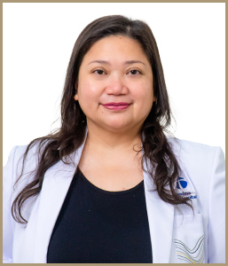
dr. Catharine Mayung Sambo, Sp.A(K)

Speciality Pediatric
Location Mandaya Royal Puri
Language Spoken Indonesia, English

Educational Background

  • Consultant Pediatrician, Growth & Development – Social Pediatrics, University of Indonesia
  • Clinical Fellow Growth & Development  – Social Pediatrics, University of Indonesia
  • Pediatrician, University of Indonesia

Courses & Training

  • IPA LEAD Workshop, International Pediatric Association -Yale Global Health Leadership Institute, Istanbul, Turkey
  • American Academy of Pediatrics National Congress and Exhibition 2019, New Orleans, USA
  • Therapeutic Use Exemption Training, World Anti-Doping Agency – Indonesia Ministry of Youth and Sport, Jakarta
  • IPA LEAD Child Health Emerging Leadership Program, International Pediatric Association, Panama
  • Media Handling Skills Training, PT Pfizer Indonesia, Jakarta
  • Protecting Children from Tobacco: Leadership Advocacy Workshop, American Academy of Pediatrics/Centers for Disease Control and Prevention, Washington DC, USA
  • Strengthening Capacity of Global Pediatric Immunization Champions in Indonesia, American Academy of Pediatrics -Indonesian Pediatric Society
  • Autism and ADHD Workshop, Indonesian Pediatric Society, Makassar
  • International Course on Facilitating Multi-stakeholder Partnership and Social Learning, Centre for Development Innovation, Wageningen University & Research, The Netherlands
  • 11th Congress, Asian Society for Pediatric Research, Japan
  • Pediatric Sleep Medicine Workshop, World Association of Sleep Medicine, Korea
  • Vaksin Anak
  • Konsultasi Kesehatan Anak
  • Konsultasi Tumbuh Kembang Anak

Primary Location

Mandaya Royal Puri

Jl. Metland Boulevard Lot. C-3 Metland Cyber City Puri, RT.001/RW.002, Parung Jaya, Kec. Karang Tengah, Kota Tangerang, Banten 15159

Appointment +6221-5092-8888
bg-left

dr. Heppy Chandra W, Sp.OT (K) Hip & Knee

Speciality Orthopedic Surgery sub. Hip & Knee, Orthopaedics & Traumatology
Location Mandaya Karawang
Language Spoken Indonesia, English

Pendidikan :

  1. Hip & Knee Subspeciality Kolegium Orthopaedi & Traumatologi Indonesia  Program Fellowship Hospital Based Hip & Knee, July 2021
  2. Orthopaedic &Traumatology Residency Program Faculty of Medicine, Airlangga University, Dr. Soetomo  General Hospital Surabaya, East Java, Indonesia, January 2017
  3. Medical Profession Education     Dr. Soetomo General Hospital Surabaya, East Java,  Indonesia, 2010
  4. Medical Education Faculty of Medicine, Airlangga University, Dr. Soetomo  General Hospital Surabaya, East Java, Indonesia  2008


Pelatihan :

  1. International Arthroscopy Training Program for Knee Arthroscopy November 2016 Seoul, South Korea
  2. AOTrauma Course – Advanced Principle of Fracture Management  December 1st – 6th 2019 Davos, Switzerland
  3. 5th Athens Shoulder Course – Cadaveric Arthroscopy Workshop 4 – 7 May 2022 Athens. Greece
  4. Shoulder and Elbow Fellowship Program March 2019 Neon Orthopaedic Clinic Seoul, South Korea
  5. 3rd Congress of Asian Pasific Federation of Societies for Reconstructive Surgery June 2016 Beijing, China
  6. 3rd AOTrauma Asia Pasific Scientific Congress & TK Experts Symposium May 2016 Chengdu, China


Penghargaan : 

  1. 1st Winner of Final Paper presentation, The 20th National Congress of Indonesian Orthopaedic  Association 2016 Jakarta, Indonesia
  2. Publication in European Journal Orthopaedic Surgery & Traumatology, The Banff Patellar Instability Instrument : Validity and Reliability of Indonesian Version
  • Arthroscopy (ACL, PCL, Meniscus) / Sport Injury
  • Arthrop;asty / Penggantian Sendi
  • Trauma & Infeksi Muskuloaketal
  • Intervensi Nyeri
  • Rekonstruksi Deformitas Tulang

Primary Location

Mandaya Karawang

Jl. Arteri Tol Karawang Barat, Teluk Jambe, Sukamakmur, Telukjambe Timur, Sukamakmur, Kec. Telukjambe Tim., Kabupaten Karawang, Jawa Barat 41361

Appointment +62267-8643-000
bg-left

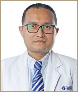
dr. Christopher Silman, Sp.Rad, Ph.D

Speciality Radiology
Location Mandaya Royal Puri
Language Spoken Indonesia, English

Dr. Christopher Silman, Sp.Rad, Ph.D., is a radiologist at Mandaya Royal Hospital Puri. He has expertise in performing various imaging procedures, including X-Ray, CT-Thorax, Mammography, CT Scan, and MRI.

Education

  • Postgraduate (Ph.D) Oita University, Japan
  • Pendidikan Dokter Spesialis Radiologi, Fakultas Kedokteran Universitas Indonesia
  • Pendidikan Dokter Umum, Fakultas Kedokteran Universitas Indonesia

Awards

  • Outstanding Reviewer Status from Canadian Association of Radiologist Journal (2018)
  • Young Investigator Award from The 6th Asian Congress of Abdominal Radiology (2017)
  • 1st Winner of the 71th Radiology National Board Exam from Indonesia Radiology National Board Exam Committee (2015)

Trainings

  • Ultrasound Masterclass: Comprehensive Musculoskeletal Ultrasound, USGH 2024
  • BIE Mammography Bootcamp with AI 2024, BIEDX 2024
  • Intensive Course and Hands-on Training in Basic Musculoskeletal Ultrasound, Indonesian Society of Ultrasound Medicine 2023
  • Prostate MRI Teaching Course 2-23 (2nd SGH PROMT)
  • Angkle, Foot, and Hip MRI Guided Mini-Fellowship and Workshop, SingHealth Academic Medical Center 2023
  • Knee and Shoulder MRI Guided Mini-Fellowship and Workshop, RADEDASIA 2022

https://www.researchgate.net/scientific-contributions/Christopher-Silman-2120459417

  • Rontgen Dada
  • Rontgen Abdomen
  • Rontgen Tulang dan Sendi
  • USG Abdomen
  • USG Doppler
  • CT-Scan Kepala
  • CT-Scan Abdomen dan Pelvis
  • CT Angiography
  • MRI Otak
  • MRI Jantung
  • MRI Muskuloskeletal
  • Fluoroskopi
  • Skrining Kanker
  • Skrining Osteoporosis

Primary Location

Mandaya Royal Puri

Jl. Metland Boulevard Lot. C-3 Metland Cyber City Puri, RT.001/RW.002, Parung Jaya, Kec. Karang Tengah, Kota Tangerang, Banten 15159

Appointment +6221-5092-8888

Patient Satisfaction Reviews

Overall
Rating:(average: 5 out of 5. Total: 1)

1 Patient Comments

  • (5/5)

    Reviewed on Jan 31, 2025

    Doctor is very helpful, informative and explain every details clearly

bg-left

drg. Claudia Ang

Speciality Dentistry
Location Mandaya Royal Puri
Language Spoken Indonesia

Educational Background

  • Dokter Umum, Universitas Trisakti

Courses & Training

  • Forum Kursus “Updating in Dentistry Life Long Learning for Better Quality of Life”
  • Oral Medicine for General Practitioners and students
  • Basic life Support Training
  • Cabut Gigi
  • Foto Panoramik Gigi
  • Pembersihan Gigi
  • Pemeriksaan dan Konsultasi Perawatan Gigi
  • Pengobatan Gigi Sensitif
  • Rontgen Gigi
  • Scaling Gigi
  • Tambal Gigi

Primary Location

Mandaya Royal Puri

Jl. Metland Boulevard Lot. C-3 Metland Cyber City Puri, RT.001/RW.002, Parung Jaya, Kec. Karang Tengah, Kota Tangerang, Banten 15159

Appointment +6221-5092-8888
Need Help? Chat with us!
Start a Conversation
Hi! Click one of our members below to chat on WhatsApp
We usually reply in a few minutes