


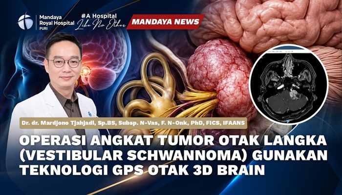
Vestibular schwannoma is a tumor that develops on the main nerve connecting the inner ear to the brain. This type of tumor is benign, meaning it is not cancerous and does not spread to other parts of the body. The affected nerve is called the vestibulocochlear nerve, which is located on both sides of the head.
The vestibulocochlear nerve plays an important role in maintaining balance and hearing. As a vestibular schwannoma grows, it can interfere with the function of this nerve and cause various symptoms, including hearing loss, ringing in the ears (tinnitus), and balance problems.
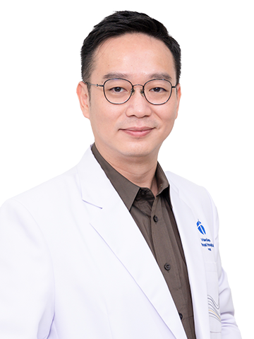
Recently, Dr. dr. Mardjono Joy Tjahjadi, Sp.BS, Subsp. N-Vas, F. N-Onk, PhD, FICS, also known as Dr. Joy, a neurosurgeon at Mandaya Royal Hospital Puri, successfully removed a vestibular schwannoma brain tumor. Approximately two days after surgery, the patient was already able to resume almost normal activities.
“There was no pain, and I can walk without feeling unsteady anymore. It feels much better,” the patient said.

Contents
Dr. Joy Explains Vestibular Schwannoma
According to Dr. Joy, vestibular schwannoma is a type of brain tumor that can develop near the brainstem and potentially cause serious problems if it continues to grow.
“Vestibular schwannoma is a type of brain tumor located near the brainstem. If the tumor becomes large, it can cause brainstem dysfunction, including paralysis, difficulty swallowing, difficulty speaking, and it can even affect the patient’s consciousness.”
“If it presses against the cerebrospinal fluid pathways, it can also cause blindness. This is a very dangerous tumor that requires medical treatment.”
In its early stages, vestibular schwannoma may not cause obvious symptoms. Symptoms usually develop gradually and can vary from one patient to another. Some symptoms that may occur include:
- Hearing loss, which usually affects only one ear.
- Tinnitus, which is the perception of ringing or other sounds originating inside the ear.
- Vertigo, a sensation that the body or surroundings are moving or spinning.
When a vestibular schwannoma becomes large, other symptoms may also develop, including:
- Persistent headaches.
- Blurred or double vision, which may occur temporarily.
- Numbness, pain, or weakness on one side of the face.
- Impaired coordination of the limbs (ataxia) on one side of the body.
- Changes in the voice or difficulty swallowing (dysphagia).
Read also: Understanding Brain Tumors: Types, Causes, Symptoms, and Treatment
Vestibular Schwannoma Brain Tumor Successfully Treated by Dr. Joy at Mandaya Royal Hospital Puri
At Mandaya Royal Hospital Puri, vestibular schwannoma removal is supported by advanced medical technologies designed to help doctors identify the location and boundaries of the tumor, as well as important surrounding nerve structures. Several technologies and surgical techniques used in the procedure include:
-
3D Brain Neuronavigation or Brain GPS
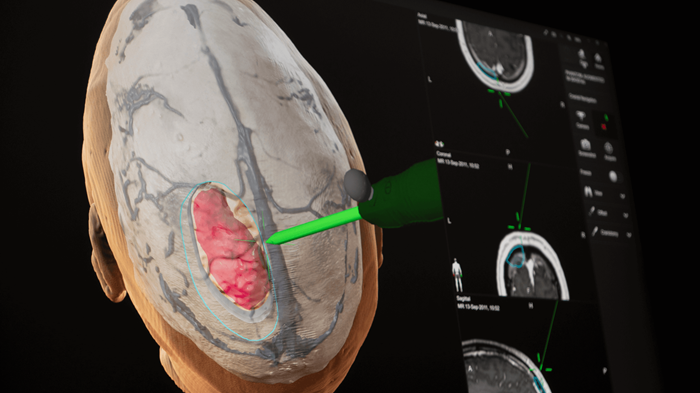
This technology helps doctors determine the location of the tumor and guides the surgical procedure more accurately based on the patient’s brain anatomy.
-
KINEVO 900S Microscope Integrated with 3D Brain Neuronavigation
The surgical microscope provides high-magnification and highly detailed visualization of the surgical area. Its integration with the neuronavigation system helps guide the surgeon throughout the tumor removal procedure.
-
Intraoperative Neuromonitoring (IONM)
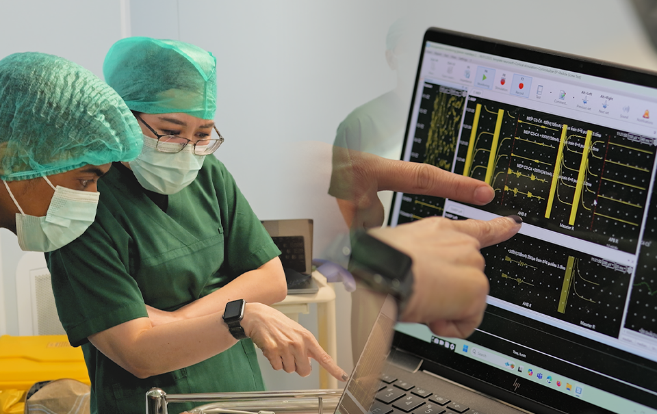
IONM is used to monitor nerve function during surgery. This monitoring allows doctors to assess the condition of important nerves in real time throughout the tumor removal procedure.
-
Microsurgical Technique
Microsurgery allows doctors to remove the tumor using high-resolution visualization and specialized instruments, with the goal of removing the tumor while preserving important surrounding structures.
Dr. Joy explained that the use of these technologies helps the medical team perform tumor removal in a more targeted manner while continuously monitoring nerve function during the procedure.
“Therefore, we perform a medical procedure called microsurgical tumor removal, guided by 3D Brain Neuronavigation and a microscope system. This allows us to clearly demarcate the tumor boundaries and then remove the tumor safely. Most importantly, we have an Intraoperative Neuromonitoring system that allows us to identify cranial nerves VII, VIII, IX, and V while removing the tumor. This helps ensure that all of these nerve functions remain stable and safe after surgery.”
Two Days After Surgery, the Patient Was Already Able to Resume Almost Normal Activities
One of the main concerns following brain tumor surgery is the patient’s recovery process. In this case, on the second day after surgery, the patient was already able to perform almost normal daily activities. This allowed the patient to be discharged on the third day after surgery.
The patient also experienced positive changes following the procedure. The patient said:
“There was no pain, and I can walk without feeling unsteady anymore. It feels much better,” the patient said.
Profile and Expertise of Dr. Joy, Brain Tumor Neurosurgeon at Mandaya Royal Hospital Puri
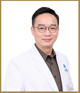
Dr. dr. Mardjono Joy Tjahjadi, Sp.BS, Subsp. N-Vas, F. N-Onk, PhD, FICS, IFAANS, commonly known as Dr. Joy, is a neurosurgeon with special expertise in vascular neurosurgery, particularly in the treatment of conditions involving the blood vessels of the brain. He completed his doctoral studies at the University of Helsinki, Finland, with a research focus on brain aneurysms, including the scientific aspects of the condition and the development of clinical treatment strategies.
In addition to treating patients, Dr. Joy is also actively involved in academic and educational activities. He is the author of Memahami Aneurisma Otak (Understanding Brain Aneurysms), a book that has been published in two editions. The book provides a comprehensive discussion of brain aneurysms, covering their causes, disease mechanisms, diagnostic processes, and available treatment options.
In his clinical practice, Dr. Joy treats various neurosurgical conditions, including:
- Brain aneurysms
- Cerebral blood vessel narrowing or stenosis
- Brain hemorrhage
- Brain tumors
- Brain cancer
With extensive clinical experience, an academic background, and training at leading neurosurgical centers around the world, Dr. Joy is experienced in managing a wide range of complex neurosurgical cases. His expertise includes the treatment of cerebral vascular disorders as well as various types of brain tumors that require highly precise surgical intervention.
Dr. Joy’s Practice Schedule at Mandaya Royal Hospital Puri
Dr. dr. Mardjono Joy Tjahjadi, Sp.BS, Subsp. N-Vas, F. N-Onk, PhD, FICS, IFAANS is available for consultation at Mandaya Royal Hospital Puri on:
- Monday: By appointment
- Tuesday: 2:00 PM–5:00 PM WIB
- Wednesday: By appointment
- Thursday: 8:00 AM–12:00 PM WIB
- Friday: 2:00 PM–5:00 PM WIB
- Saturday: 8:00 AM–12:00 PM WIB
To make your visit to Mandaya Royal Hospital Puri more convenient, use the Chat via WhatsApp, Book an Appointment, or the Care Dokter app, which can be downloaded from Google Play and the App Store. The app can also be used to check queue numbers and access other relevant information.
The information provided on this page is intended for educational purposes and general information only and does not represent the full range of medical services that may be provided by each doctor. To determine the appropriate treatment for your specific health condition, consultation with the relevant doctor is recommended.
If you have any questions, suggestions, or require further information, please contact our call center at 0811-1900-2000.

References:
- Mayo Clinic. Acoustic Neuroma: Symptoms and Causes. (https://www.mayoclinic.org/diseases-conditions/acoustic-neuroma/symptoms-causes/syc-20356127). Accessed August 10, 2026.
- NHS. Acoustic Neuroma. (https://www.nhs.uk/conditions/acoustic-neuroma/). Accessed August 10, 2026.