Brain tumors are serious medical conditions that require highly careful management, as the brain plays a crucial role in controlling various body functions. From movement and speech to vision, all are regulated by this vital organ. Therefore, brain tumor surgery relies not only on the surgeon’s expertise but also on advanced technology to ensure the procedure is performed safely and with high precision.
Currently, brain tumor surgeries performed by Dr. Joy at Mandaya Royal Puri Hospital are supported by 3D real-time neuronavigation technology. Often referred to as “brain GPS,” this system helps surgeons accurately locate tumors and determine the most optimal surgical pathway. With this technology, tumor removal can be performed more effectively while minimizing the risk of damage to healthy brain tissue.
Dr. dr. Mardjono Joy Tjahjadi, Sp.BS, Subsp. N-Vas, F. N-Onk, PhD, FICS, IFAANS, also known as Dr. Joy, is a neurosurgeon with extensive experience in handling complex neurological cases. In certain conditions, such as tumors located in difficult-to-reach areas including trigone meningioma, neuronavigation technology plays a significant role in improving surgical precision.

Contents
Why Is 3D Neuronavigation Important in Brain Tumor Surgery?
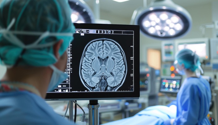
In modern neurosurgery, accuracy is a key factor in achieving successful outcomes. Surgeons need clear guidance to identify the tumor location and determine the safest surgical approach. Neuronavigation technology provides a solution by offering a real-time, three-dimensional map of the brain during surgery.
Through this system, doctors can better understand the relationship between the tumor and surrounding brain tissues. This allows for more targeted surgical procedures while helping protect critical functional areas of the brain.
Key benefits of this technology include:
- Precise identification of tumor location based on MRI or CT scan data
- Safer and more efficient surgical pathway planning
- Real-time visualization of surgical instruments during the procedure
- Reduced risk of damage to vital brain areas
- Clearer tumor boundaries for more optimal removal
This technology is particularly beneficial for tumors located deep within the brain or in hard-to-reach areas. With accurate navigation, surgical success rates can improve while reducing the likelihood of complications.
How Does 3D Neuronavigation Work?
The neuronavigation system operates through several integrated steps, from initial diagnosis to the surgical procedure:
- The patient undergoes an MRI to obtain detailed images of the brain
- MRI data is uploaded into the neuronavigation system to create a digital brain map
- The surgeon develops a comprehensive surgical plan using the system
- The system is aligned with the patient’s head position in the operating room
- The tumor location is marked as a surgical guide
- Brain structures, including the tumor and blood vessels, are displayed with color-coded visualization
- The system helps determine the safest surgical pathway
Through these steps, neuronavigation technology enhances surgical accuracy while improving patient safety during the procedure.
Patient Story: Optimal Surgical Outcome with 3D Real-Time Neuronavigation
One successful case at Mandaya Royal Puri Hospital involved Mr. GBD, a content creator known as Elder Storyteller. He underwent surgery to remove a meningioma tumor measuring approximately 4×4 cm with Dr. Joy.
His symptoms began with sudden seizures while eating with his family. After undergoing CT scan and MRI examinations, a tumor was found on the left side of the brain, pressing on nerves and affecting vision. The tumor was also suspected to be the cause of previous visual disturbances and several falls.
With the support of neuronavigation technology, approximately 99% of the tumor was successfully removed based on post-operative evaluation. His recovery was relatively fast, as he did not require ICU care and was able to sit, stand, and walk within a few days.
This case demonstrates how the combination of a surgeon’s expertise and modern technology can lead to more optimal outcomes in brain tumor treatment.
Expertise and Practice Schedule of Dr. Joy at Mandaya Royal Puri

Dr. Joy is a neurosurgeon with a subspecialty in cerebrovascular diseases. He completed his doctoral studies at the University of Helsinki, Finland, focusing on brain aneurysms.
In addition to clinical practice, he is actively involved in academic work and has authored the book “Memahami Aneurisma Otak” (Understanding Brain Aneurysms), which provides a comprehensive discussion of the condition.
In his practice, he manages various complex neurological conditions, including:
- Brain aneurysms
- Cerebral blood vessel narrowing
- Brain hemorrhage
- Brain tumors
- Brain cancer
With extensive clinical experience and international training, Dr. Joy is recognized as one of Indonesia’s leading neurosurgeons.
Dr. dr. Mardjono Joy Tjahjadi is available at Mandaya Royal Puri Hospital on:
- Monday: by appointment
- Tuesday: 14:00 – 17:00 WIB
- Wednesday: by appointment
- Thursday: 08:00 – 12:00 WIB
- Friday: 14:00 – 17:00 WIB
- Saturday: 08:00 – 12:00 WIB
To make hospital visits more convenient, patients can use WhatsApp Chat, Book Appointment services, or the Care Dokter app available on Google Play and the App Store. These services allow patients to schedule consultations, check queue numbers, and access important hospital information.
The information provided on this page is intended for general educational purposes and does not represent the full scope of medical services offered by each doctor. To ensure appropriate care based on your condition, it is recommended to consult directly with the relevant doctor.
If you have any questions, suggestions, or need further information, please contact our call center at 0811-1900-2000.