Liver cancer is a type of cancer that occurs when cells in the liver grow abnormally and form tumors. In its early stages, liver cancer often does not cause clear symptoms. However, as the disease progresses, patients may experience various complaints such as pain in the upper right abdomen, abdominal swelling, unexplained weight loss, fatigue, and yellowing of the skin and eyes. If not treated properly, this condition can interfere with liver function and significantly reduce a patient’s quality of life.
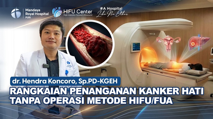
Today, advances in medical technology have enabled more modern and minimally invasive treatment options for liver cancer, one of which is High Intensity Focused Ultrasound (HIFU) or Focused Ultrasound Ablation (FUA). According to Hendra Koncoro, Sp.PD-KGEH, an internal medicine specialist and gastroenterohepatology consultant at Mandaya Royal Hospital Puri, this technology uses high-energy ultrasound waves that are focused directly on tumor tissue to destroy cancer cells without requiring surgical incisions. This method can be considered as an alternative treatment option for liver cancer in patients with certain medical conditions.

Contents
HIFU / FUA Technology for Liver Cancer Treatment
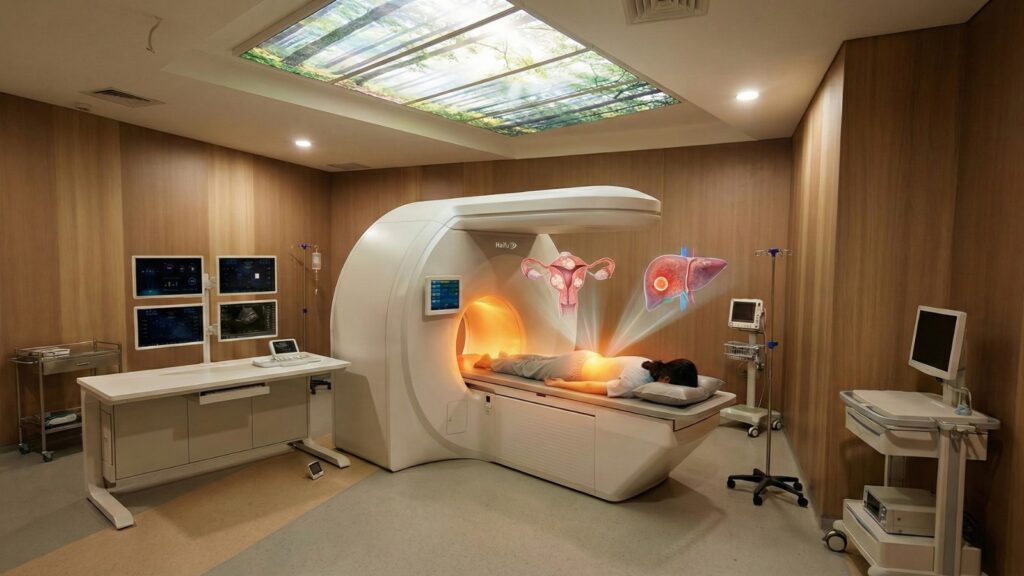
The HIFU or FUA method uses high-energy ultrasound waves that are precisely focused on the tumor area to destroy cancer tissue without the need for surgery.
According to dr. Hendra, this technology can be utilized to treat various conditions related to liver cancer, including complications that are often difficult to manage with other treatment methods.
“Currently, we are performing Focused Ultrasound Ablation procedures for malignancies in the field of hepatology, particularly liver cancer,” said dr. Hendra.
This procedure works by delivering focused ultrasound energy to the targeted tissue. The energy generates heat that can destroy cancer cells precisely while minimizing damage to surrounding healthy tissue.
Liver Cancer Case with Portal Vein Thrombus
In one of the cases handled by dr. Hendra, a 68-year-old female patient had been diagnosed with advanced-stage liver cancer since 2023. She had previously undergone various treatments, but a complication called portal vein thrombosis was still present, which is a blood clot in the vein caused by tumor growth.
According to dr. Hendra, this condition is considered a challenging complication to treat.
“Portal vein thrombus is an advanced complication of liver cancer that causes blood clots in the blood vessels due to the tumor, and it is generally very difficult to remove,” he explained.
Through HIFU or FUA technology, the ablation process can be performed directly on the area where the thrombus is located. During the procedure, the patient is placed under sedation administered by an anesthesiologist, and ultrasound energy is focused on the target area to destroy the tumor tissue causing the blockage.
Minimally Invasive Procedure with Relatively Low Risk
One of the main advantages of the HIFU / FUA method is that it is non-invasive, meaning it does not require open surgery. The procedure typically takes around two to two and a half hours.
“This procedure takes about two to two and a half hours with the patient under sedation, followed by focused ablation using ultrasound waves,” explained dr. Hendra.
Because it does not involve surgical incisions, the risk of complications is generally lower compared to conventional surgical methods. After the procedure, the patient will undergo follow-up evaluations to assess the treatment response, including imaging examinations such as MRI.
“Evaluation will be performed about two weeks after the procedure to determine whether the portal vein thrombus has decreased,” he added.
If the evaluation results show improvement, further therapies may be considered, such as immunotherapy, Transarterial Chemoembolization (TACE), or ablation using radiofrequency ablation (RFA) or microwave ablation. This integrated approach is expected to help control the disease while improving the quality of life of liver cancer patients.
Profile and Practice Schedule of dr. Hendra at Mandaya Royal Hospital Puri

dr. Hendra Koncoro, Sp.PD-KGEH is an internal medicine specialist with a subspecialty as a gastroenterohepatology consultant. He has expertise in treating various diseases related to the digestive system and liver, ranging from common conditions to more complex cases. Some of the conditions he treats include liver cancer, gallstones, fatty liver, liver nodules, liver cirrhosis, and bile duct cancer.
During his educational journey, dr. Hendra obtained his medical degree from the Atma Jaya Catholic University of Indonesia. He then continued his Master of Biomedical Science at Udayana University. After that, he pursued his Internal Medicine Specialist training at the same university before continuing his subspecialty training as a Gastroenterology and Hepatology Consultant at the University of Indonesia.
As a gastroenterohepatology consultant, dr. Hendra also provides various medical consultation and diagnostic services to help diagnose and monitor patient conditions. These services include internal medicine consultations, liver function tests, urine tests, lipid profile tests, and supporting examinations such as CT Scan, MRI Scan, and kidney function tests.
With a combination of strong academic background and extensive clinical experience, dr. Hendra is committed to providing accurate diagnosis and optimal treatment for patients with digestive system and liver disorders.
Patients can meet dr. Hendra Koncoro at Mandaya Royal Hospital Puri on the following schedule:
- Wednesday: 10.30 – 12.30 WIB
- Saturday: 10.30 – 12.30 WIB
To make your visit easier, you can use the WhatsApp Chat, Book Appointment, or the Care Dokter application, available on Google Play and App Store, to schedule visits, check queue numbers, and obtain other important information before coming to Mandaya Royal Hospital Puri.