A liver tumor is an abnormal growth of tissue in the liver that can be benign (non-cancerous) or malignant (cancerous). Benign liver tumors are relatively common and usually cause no symptoms, often discovered incidentally during ultrasound, CT scan, or MRI. Examples include hepatocellular adenoma, which rarely turns cancerous, and hemangioma, which typically requires no special treatment.
Malignant liver tumors, on the other hand, can originate directly from the liver (primary liver cancer) or spread from other organs (metastatic liver cancer). Most liver cancer cases are metastatic. This is why early detection and diagnosis are crucial to determine the type of tumor and select the right treatment.
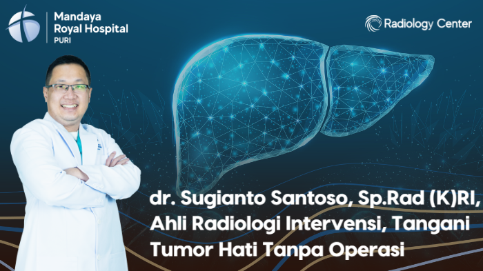
In many cases, doctors perform surgery to remove liver tumors. However, there are also minimally invasive, non-surgical methods that can be recommended, including Radiofrequency Ablation (RFA), Microwave Ablation (MWA), and Transarterial Chemoembolization (TACE). At Mandaya Royal Hospital Puri, these advanced procedures are performed by consultant interventional radiologist dr. Sugianto Santoso, Sp.Rad (K)RI.
Contents
Understanding RFA, MWA, and TACE in Liver Tumor Treatment: dr. Sugi’s Expertise1. Radiofrequency Ablation (RFA)
RFA is a modern technique for destroying liver tumors, whether primary or metastatic, especially when surgery is not an option.
During this procedure, the doctor uses ultrasound guidance to insert a special needle (probe) directly into the tumor. The probe delivers high-frequency electrical currents that generate heat, which burns and destroys tumor cells without damaging healthy surrounding liver tissue.
Benefits of RFA for liver tumors:
- Clinically proven: Widely used in thousands of patients in the U.S. and Europe.
- Minimally invasive: Requires only a needle, leaving smaller wounds and enabling faster recovery.
- Safe and effective: Precisely targets tumor cells without harming healthy liver tissue.
- Short recovery time: Patients can usually go home sooner compared to major surgery.
- Alternative for inoperable tumors: A solution for patients whose tumors cannot be surgically removed.
2. Microwave Ablation (MWA)
MWA uses heat from microwave energy to destroy liver tumors. A thin antenna is inserted into the tumor under CT or ultrasound guidance, emitting microwaves that generate high heat, destroying tumor tissue in as little as 10 minutes.
Advantages of MWA for liver tumors:
- Faster procedure: Works quicker than RFA, reducing anesthesia duration.
- Multiple tumors treated simultaneously: Allows ablation of several liver tumors in one session.
- Effective for larger tumors: Can treat bigger tumors compared to RFA.
3. Transarterial Chemoembolization (TACE)
TACE is a minimally invasive, non-surgical treatment combining chemotherapy delivery with embolization (blocking blood supply). Typically performed by interventional radiologists like dr. Sugi, it is especially useful for liver cancer.
During TACE, chemotherapy drugs are injected directly into the artery feeding the tumor, followed by embolic agents that block blood flow. This traps the drug inside the tumor longer while cutting off its blood supply, leading to more effective tumor cell destruction.
Benefits of TACE:
- Stops or shrinks tumors: Effective in about two-thirds of liver cancer cases.
- Lasting results: Benefits usually last 10–14 months; repeatable if cancer recurs.
- Combinable with other therapies: Can be paired with ablation, chemotherapy, or radiotherapy depending on tumor size and location.
- Preserves liver function: Slows tumor growth, reducing the risk of liver failure.
- Improves quality of life: Helps patients maintain better overall well-being during treatment.
Ultrasound/CT-Guided Liver Biopsy: Diagnosing Liver Tumors
To determine whether a liver tumor is benign or malignant, dr. Sugi performs ultrasound- or CT-guided liver biopsy at Mandaya Royal Hospital Puri.
- Ultrasound-guided biopsy: A small liver tissue sample is taken for lab analysis. Ultrasound helps doctors precisely locate suspicious areas. The procedure is safe, effective, outpatient-based, and results are typically available within a few days.
- CT-guided biopsy: Uses advanced X-ray imaging to create 3D scans of the liver, helping doctors identify exact areas for sampling. While ultrasound is often preferred due to its accessibility and no radiation, CT can be more accurate when ultrasound fails to detect problematic tissue.
About dr. Sugianto Santoso, Sp.Rad (K)RI

dr. Sugianto Santoso, Sp.Rad (K)RI is a consultant interventional radiologist experienced in performing various minimally invasive procedures for diagnosing and treating conditions across multiple organs.
With radiology imaging guidance, he manages conditions such as thyroid nodules, liver abscesses, and vascular disorders, including brain aneurysms. His advanced skills include procedures like brain DSA, DSA Coiling, DSA Flow Diverter, and DSA Stenting.
- Medical Education:General Medicine at Maranatha Christian University, Bandung.
- Specialization:Radiology at the University of Indonesia.
Clinic Hours at Mandaya Royal Hospital Puri
You can meet dr. Sugianto Santoso, Sp.Rad (K)RI at Mandaya Royal Hospital Puri on:
- Tuesday: 16:00 – 20:00 WIB
- Thursday: 16:00 – 20:00 WIB
For easier access, book your appointment via WhatsApp Chat, the Book Appointment feature, or the Care Dokter App (available on Google Play and App Store) to check queue numbers and get complete hospital information.


