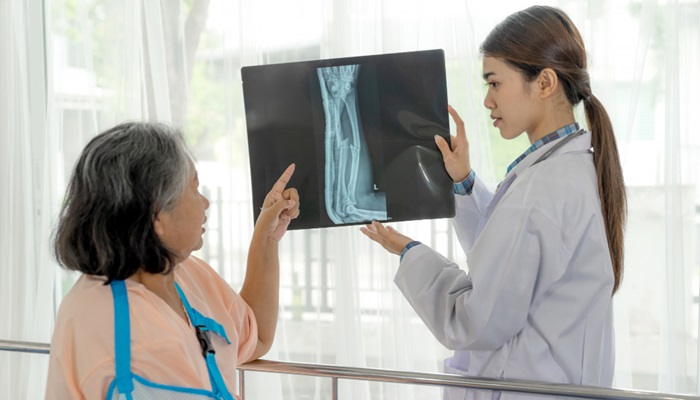
MRI for bones and joints is a scan using magnetic waves and radio frequencies to observe and examine the condition of bones, joints, and other soft tissues like cartilage, muscles, and tendons. This examination can be conducted on any part of the body with bones and joints that are suspected to have issues.
Compared to other imaging technologies, such as X-rays and CT scans, MRI can produce clearer images without exposure to radiation.
MRI for bones and joints can be divided into individual exams, based on the area to be examined, such as knee MRI, pelvic bone MRI, spinal MRI (cervical or lumbar), or shoulder MRI.

Contents
Benefits of MRI for Bones and Joints
The main benefit of MRI for bones and joints is to examine bones, joints, and other soft tissues like cartilage, muscles, and tendons. Typically, an MRI for bones and joints is performed post-injury or when there are abnormal conditions affecting the bones and joints, such as tumors, bone deformities, and birth defects.
Some conditions that might require an MRI for bones and joints include:
- Injuries, such as fractures or tears in tendons, ligaments, or cartilage
- Structural changes in bones due to aging
- Bone infections, such as osteomyelitis
- Bone tumors or cancer
- Congenital diseases
- Osteonecrosis (death of bone cells due to lack of blood supply to the area)
- Bone marrow diseases
- Degenerative joint issues, such as arthritis
- Pinched nerves or herniated discs (HNP) in the spine
- Post-surgery evaluations of specific bone or joint procedures
Preparing for MRI for Bones and Joints
Like other MRI procedures, an MRI for bones and joints has certain considerations to be aware of before scheduling the scan.
MRI uses a large magnetic field. Therefore, if you have any metal health devices implanted in your body or have undergone surgery for metal plates or screws, you must inform your doctor.
Metallic materials can interact with the strong magnetic field, potentially damaging the health devices or causing inaccurate results.
Inform your doctor if you have or use any of the following:
- Pacemaker
- Aneurysm clips in the skull
- Cochlear implants
- Certain organ prostheses
- Neurostimulators
- Some types of IUDs
- Tattoos or piercings on the body
- Metal objects inside the body, such as bullets, surgical clips, plates, or wire mesh.
Additionally, if you suffer from claustrophobia, inform the staff. The doctor or technician will provide you with light sedation to help you relax.
Procedure
Here are the steps for undergoing an MRI for bones and joints:
- You will be asked to change into a special MRI gown and remove all metal accessories, including underwire bras, hairpins, and glasses.
- If the MRI is performed with contrast, the technician will insert an IV line for the injection of the contrast material.
- You will be asked to lie down on the examination table. You may be given earplugs or headphones to listen to music and block out the noise from the MRI machine. The table will then move into the MRI machine.
- During the exam, you will need to lie still to ensure clear images. You may be asked to hold your breath for a few seconds during the scan.
- If contrast is used, you may feel a cold or uncomfortable sensation at the injection site, usually in the hand or arm.
- After the exam, the table will be moved out of the machine. If contrast was used, the technician will remove the needle.
MRI procedures usually take between 30 to 60 minutes, depending on the area of the bone or joint being examined. In some cases, a radiofrequency coil may be used to gather magnetic waves and produce clearer images.
Typically, MRIs, including those for bones and joints, are outpatient procedures. This means you can go home immediately after the scan. If you receive light sedation, you may need to wait 1-2 hours for the effects to wear off.
Risks of MRI for Bones and Joints
MRI for bones and joints does not use radiation, so there is no risk of radiation exposure during the scan. However, you must inform the staff about any metal health devices to avoid the risk of damage.
Other risks may include allergic reactions to the contrast material and a metallic taste in your mouth. Bruising at the injection site can also occur, but this is usually mild.
To determine the appropriate type of MRI for bones and joints and the right timing, consult with your doctor. An orthopedic specialist can help determine the correct area for the MRI.
You can also visit Preventive Health Care at RS Mandaya Royal for a bone and joint MRI. Schedule an appointment via WhatsApp, Book Appointment, or download the Care Dokter app on Google Play and the App Store.
Treatment of Osteoarthritis (OA) or Joint Degeneration by Dr. Jecky Chandra, Sp.OT, M.Kes