Brain tumor surgery is a medical procedure that requires an extremely high level of precision. The brain contains many vital structures, such as nerves and blood vessels, that play essential roles in various bodily functions. Therefore, surgical procedures in this area must be carefully planned and supported by advanced technology.
To improve safety and accuracy in brain tumor surgery, RS Mandaya Royal Puri provides 3D brain mapping technology, also known as neuronavigation, from Brainlab. This technology helps neurosurgeons clearly visualize the position of the tumor through a three-dimensional display on the screen during surgery. With the support of this system, doctors can determine a safer surgical pathway so that the tumor removal process can be performed with greater precision.
At RS Mandaya Royal Puri, this technology is used by Dr. dr. Mardjono Joy Tjahjadi, Sp.BS, Subsp. N-Vas, F. N-Onk, PhD, FICS, IFAANS, better known as Dr. Joy. He is a neurosurgeon experienced in handling various complex neurological cases. In several cases, including rare brain tumors such as trigone meningioma, this brain mapping technology helps doctors perform surgery more accurately, allowing tumors to be removed in a safer and more targeted manner.

Contents
How 3D Brain Mapping Technology Works During Brain Tumor Surgery
Neuronavigation technology works by combining medical imaging results with a digital navigation system used during surgery. Through this system, doctors can view brain structures in detail and determine the safest pathway to reach the tumor. Below are the stages of using this technology in brain tumor surgery.
1. Brain MRI Examination
The first stage begins with a brain MRI examination. This imaging test aims to obtain a highly detailed view of the patient’s brain condition, including the tumor’s location, size, and its relationship with surrounding brain tissue.
2. Integration of MRI Data into the Navigation System
After the examination is completed, the MRI data is entered into the Brainlab neuronavigation system. The system then processes the data digitally and displays it in the form of a three-dimensional brain map.
3. Surgical Planning
The neurosurgeon then uses the system to plan the surgical strategy. At this stage, the doctor determines the safest surgical approach to reach the tumor.
4. Alignment with the Patient’s Head Position
In the operating room, the digital brain map is adjusted to match the patient’s actual head position. This process is called registration, which aligns the digital map with the patient’s anatomical structure in real time.
5. Tumor Location Marking
The doctor then marks the tumor location on the navigation system screen. This marking serves as the main guide during the surgical procedure.
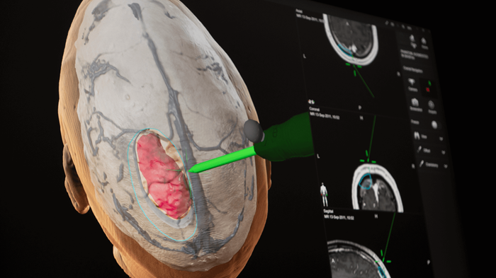
6. Visualization of Brain Structures
The system screen displays detailed images of the brain, including the tumor and surrounding blood vessels. These structures are usually shown in different colors to make them easier to identify during surgery.
7. Determining the Safest Surgical Path
With the help of this navigation system, the doctor can determine the safest path to reach the tumor. This technology helps minimize the risk of damage to critical brain tissue while improving the accuracy of tumor removal.
Benefits of Brain GPS Technology in Tumor Surgery
In modern neurosurgery, accuracy is a key factor that determines surgical success. Doctors need clear guidance to identify the tumor’s position while preserving healthy brain tissue. For this reason, RS Mandaya Royal Puri uses neuronavigation technology, often referred to as brain GPS.
This technology can map brain structures in three dimensions and display them in real time during surgery. As a result, doctors can clearly see the relationship between the tumor and the surrounding brain tissue.
Some of the main benefits of this technology in brain tumor surgery include:
- Helping determine the tumor location more precisely based on MRI or CT scan results
- Assisting doctors in planning the safest surgical pathway
- Displaying the position of surgical instruments in real time on the digital brain map
- Reducing the risk of injury to important brain areas such as those responsible for movement, speech, and vision
- Helping doctors clearly identify tumor boundaries so the chances of maximum tumor removal increase
This 3D brain mapping technology is particularly useful for tumors located deep within the brain or in difficult-to-reach areas, such as trigone meningioma. With precise navigation, surgical procedures can be performed with better safety and higher success rates.
Patient Story: Rare Brain Tumor Surgery Handled with 3D Brain Mapping Technology by Dr. Joy
One example of successful brain tumor surgery at RS Mandaya Royal Puri was experienced by Mr. GBD, a content creator known on social media as Elder Storyteller. He underwent surgery to remove a 4 × 4 cm meningioma brain tumor with Dr. Joy.
Initially, Mr. GBD experienced sudden seizures while having a meal with his family. After undergoing CT scan and MRI examinations, doctors discovered a tumor on the left side of his brain that was pressing on nerves and affecting his vision.
The tumor was also suspected to be the cause of several symptoms he had previously experienced, including visual disturbances and several sudden falls.
Through surgery supported by neuronavigation or brain GPS technology, the tumor was successfully removed by approximately 99%, based on postoperative CT scan and MRI results. Mr. GBD’s recovery process was also relatively fast. After surgery, he did not need ICU care and was already able to sit, stand, and walk within a few days.
This story demonstrates how the combination of a neurosurgeon’s expertise and modern medical technology can improve the success of brain tumor surgery while also accelerating the patient’s recovery process.
Profile and Practice Schedule of Dr. Joy at RS Mandaya Royal Puri

Dr. Joy is a neurosurgeon with a subspecialty in cerebrovascular diseases. He earned his doctoral degree from the University of Helsinki, Finland, with a research focus on brain aneurysms and the development of clinical approaches for their management.
In addition to actively treating patients, Dr. Joy also contributes to the advancement of medical science through academic activities. He is the author of the book “Memahami Aneurisma Otak” (Understanding Brain Aneurysms), which has been published in two editions. The book discusses brain aneurysms comprehensively, including their causes, disease mechanisms, diagnostic methods, and treatment options.
In his clinical practice, Dr. Joy has extensive experience in treating various neurological conditions, including:
- Brain aneurysm
- Narrowing of brain blood vessels (stenosis)
- Brain hemorrhage
- Brain tumors
- Brain cancer
With extensive clinical experience, a strong academic background, and international training at leading neurosurgery centers around the world, Dr. Joy is recognized as one of the experienced neurosurgeons in Indonesia.
For patients who wish to consult, the practice schedule of Dr. Joy at RS Mandaya Royal Puri is as follows:
- Monday: by appointment
- Tuesday: 14:00 – 17:00 WIB
- Wednesday: by appointment
- Thursday: 08:00 – 12:00 WIB
- Friday: 14:00 – 17:00 WIB
- Saturday: 08:00 – 12:00 WIB
To make visits more convenient, patients can use several digital services provided by RS Mandaya Royal Puri, including WhatsApp Chat, Book Appointment, or the Care Dokter application available on Google Play and the App Store. Through these services, patients can schedule consultations, check queue numbers, and obtain various important information related to hospital services.